

Abstract
Introduction
Immune thrombocytopenia (ITP) is an autoimmune disease in which autoreactive T and B cells are activated by platelet autoantigens resulting in immune-mediated platelet destruction and/or suppression of platelet production. The binding of programmed death 1 (PD-1) to its ligands PD-L1 and PD-L2 on antigen-presenting cells turns off autoreactive T cells and induces peripheral tolerance. Aberrant PD-1/PD-L signalling could result in a breakdown of peripheral tolerance and lead to autoimmune diseases.
Methods
Thirty-four patients with primary active ITP who were diagnosed and/or followed up and 26 healthy controls were enrolled in this study. Platelet counts in all ITP patients were less than 30×109 /L at sampling. They had not been treated with any immunosuppressive agents for at least one week prior to sampling for this study.
To determine the role of the PD-1/PD-L signalling pathway in ITP, we detected PD-1 expression on T cells and PD-L expression on dendritic cells (DCs) in both ITP patients with active disease and healthy controls by flow cytometry. To investigate the effects of PD-L1-Fc fusion protein (PD-L1-Fc) on T cells, PBMCs from ITP patients and healthy controls with autologous platelets were cultured with soluble anti-CD3 monoclonal antibody (mAb) and anti-CD28 mAb in the presence (PD-L1-Fc+ group) or absence (PD-L1-Fc- group) of the PD-L1-Fc (0.5 μg/mL) at 37 ˚C with 5% CO2 for 4 days. The cells were harvested and stained for flow cytometry to detect the apoptosis, activation and proliferation of T cells. IL-2 and IFN-γ levels in the co-culture supernatant were assayed by ELISA.
Results
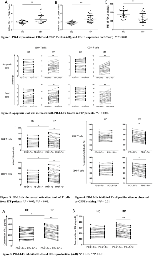
Enhanced PD-1 expression on T cells and decreased PD-L1 expression on mDCs in ITP patients
We found that PD-1 mean fluorescence intensities (MFIs) increased in CD4+ cells (P < 0.01) and in CD8+ cells (P < 0.01) from ITP patients compared with those from healthy controls. However, PD-L1 expression on monocyte-derived DCs was lower in patients with active ITP than in healthy controls (P < 0.01, Figure 1).
PD-L1-Fc promoted T cell apoptosis
Annexin V-FITC and propidium iodide (PI) were used to detect T cell apoptosis. The percentage of apoptotic cells and dead cells were analysed to determine T cell apoptosis levels. In ITP patients, the percentage of apoptotic cells and dead cells were higher in PD-L1-Fc+ group than in the PD-L1-Fc- group (P < 0.01 for both CD4+ and CD8+ T cells). However, we found no significant difference in apoptosis between PD-L1-Fc- and PD-L1-Fc+ groups in healthy controls (Figure 2).
PD-L1-Fc inhibited T cell activation and proliferation
CD25 MFIs were analysed to determine the activation level of cocultured T lymphocytes. Compared with healthy controls, CD25 expression on CD4+ and CD8+ T cells was significantly increased in ITP patients (P < 0.05 for both CD4+ and CD8+ T cells). These results suggest that ITP patients had more activated CD4+ and CD8+ T cells than in healthy controls. PD-L1-Fc significantly inhibited T cell activation in ITP patients (P < 0.01, n=34) but not in healthy controls (P = 0.0834 in CD4+ T cells, P = 0.6834 in CD8+ T cells, n=26, Figure 3).
To analyse proliferation, the frequency of divided cells was calculated according to the loss of CFSE fluorescence intensity. PD-L1-Fc inhibited proliferation of T cells from ITP patients (P < 0.01 in both CD4+ T and CD8+ T cells, n=34, Figure 4). We found no significant difference in proliferation in T cells from healthy controls (CD4+ T cells P = 0.9758, CD8+ T cells P = 0.5658, n=26).
PD-L1-Fc inhibited IL-2 and IFN-γ production
IL-2 secretion was higher in ITP than in healthy controls in the absence of PD-L1-Fc (P = 0.0308, Figure 5). PD-L1-Fc significantly inhibited IL-2 and IFN-γ production in ITP patients. IL-2 and IFN-γ levels were lower in the PD-L1-Fc+ group than in the PD-L1-Fc- group in ITP (P < 0.01 for both IL-2 and IFN-γ, n=34). However, we did not detect a significant difference in secretion of IL-2 (P = 0.2016, n=26) or IFN-γ (P = 0.2989, n=26) in healthy controls.
Conclusions
In summary, our study suggests that the aberrant PD-1/PD-L negative costimulatory pathway may play a role in ITP. Enhancing PD-1/PD-L signalling might be a promising therapeutic approach for ITP by promoting T cell apoptosis, inhibiting T cell activation and proliferation, and reducing secretion of inflammatory factors.
No relevant conflicts of interest to declare.

This feature is available to Subscribers Only
Sign In or Create an Account Close Modal